Ablation means destroying tissue and cryo means cold or freezing.
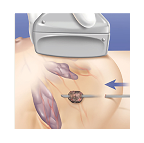
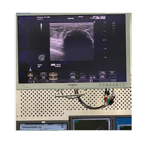
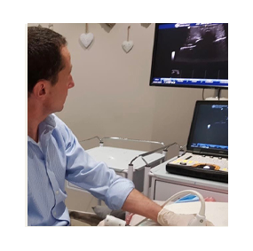
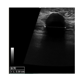
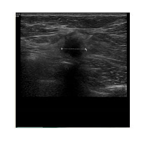
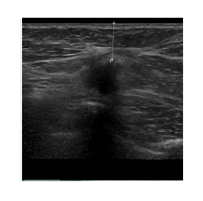
The freezing is achieved with liquid nitrogen which flows through a probe (needle.) The needle is inserted into the tumor under ultrasound guidance. This allows us to position it very precisely, ensuring that the tumor is at the centre of the freezing zone.
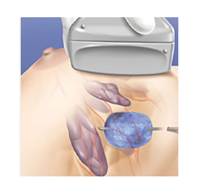
This creates a large ball of ice that engulfs the tumor. By alternating freeze and thaw cycles, the cells in the tumor are destroyed. By the end of the procedure there should be no viable cancer cells left. The progress of ice ball development is closely monitored throughout the procedure to ensure safety.
The procedure is performed after the administration of local anesthetic. The skin and deeper tissues are completely numbed prior to the introduction of the cryoablation needle.
It is therefore a painless procedure. Cryoablation of a breast tumor takes 30 – 40 minutes. Patients can go home soon thereafter.
Follow up studies with mammograms, ultrasound and MRI confirm the absence of any residual breast cancer.
Since cryoablation involves tissue destruction, patients can expect to develop bruising of the breast that lasts usually for one to two weeks. Post procedure pain is usually mild and can be controlled with anti-inflammatories and paracetamol.
A palpable lump remains at the tumor site after cryoablation. This is scar tissue that forms after tumor destruction. It will resolve within 6 to 12 months.
The purpose of cryoablation is to offer an alternative to surgery for selected breast cancers.
INTERNATIONAL
International experience and Clinical Trials
Cryoablation is gaining worldwide recognition as a minimally invasive and highly successful alternative to surgery for small breast cancers. Multiple clinical trials over the last 10 years have proven the effectiveness of the procedure, and shown extremely low recurrence rates after treatment.
The most recent trial – ICE 3 – was published in 2024 and confirmed an absence of recurrent disease in 96% of patients after 5 years.
The liquid nitrogen never leaves the needle, but it causes the tissue around the needle tip to freeze rapidly.
In 2017, the Parklane Mammography department became the first unit to be accredited by the America College of Radiologists.
Dr. Schoub is an active participant in several multidisciplinary breast cancer teams across Johannesburg, collaborating closely with leading surgeons and oncologists. In 2020 Dr Schoub, established the first cryoablation facility in South Africa. In the last 4 years more than 75 cryoablation procedures have been successfully performed in the department.
In the last 4 years more than 75 cryoablation procedures have been successfully performed in the department.
PROCEDURE DIAGRAM
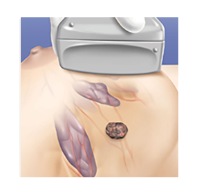
Ultrasound imaging is used to locate the lesion. The breast is prepared and local anesthesia is given
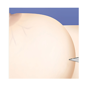
A 3mm incision (about 1/8 inch) is made in a cosmetically and technically appropriate location
Using ultrasound guidance, a visica cryoprobe is positioned in the center of the lesion
The cycle is activated and an iceball forms around the tumor. The freezing temperatures destroy the tumor tissue
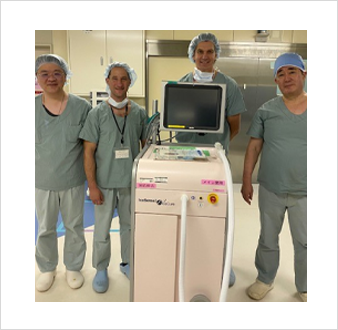
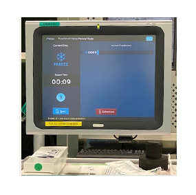
The Icecure Prosense console
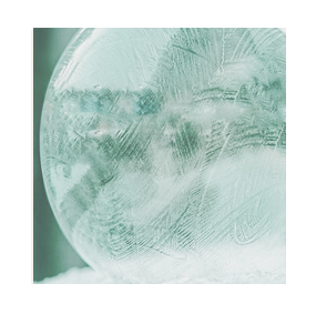
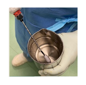
Testing – the ice ball forms around the probe
Freeze and thaw cycles are used sequentially to destroy the tumour